
Ankle Cartilage Restoration
Ankle Cartilage ‘Restoration’……..
Losing cartilage does not necessarily mean losing the joint
An ankle injury can sometimes leave behind more than a sprain.
A significant ankle sprain, fracture or sporting injury can damage the smooth cartilage that covers the surface of the ankle joint. In some patients, a small area of cartilage and the underlying bone is injured. This is known as an osteochondral lesion of the talus, or OLT.
For many years, finding cartilage damage in the ankle could feel rather final.
Fortunately, that is no longer always the case.
We cannot simply make damaged cartilage completely normal again. However, for the right type of cartilage injury, modern joint-preserving techniques can encourage the ankle to form new repair tissue and restore a more functional joint surface.
One of the techniques I use for selected patients is AMIC, or Autologous Matrix-Induced Chondrogenesis, often using a special collagen membrane.
The aim is simple:
repair what we can, preserve the ankle joint and keep you moving.
What is cartilage?
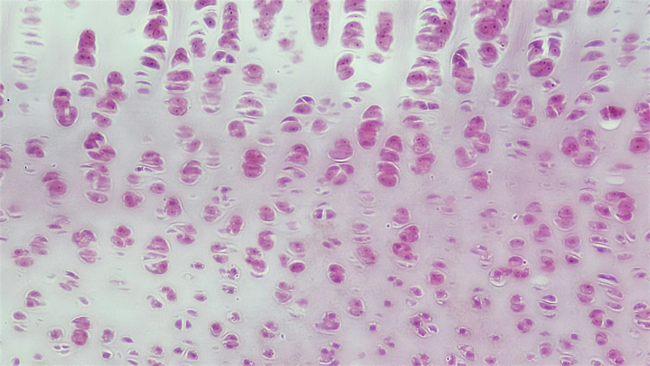
Articular cartilage is the smooth, slippery surface covering the ends of the bones within a joint. -hyaline cartilage’.
In the ankle, cartilage covers the top of the talus and the bottom of the tibia. Healthy cartilage allows these surfaces to glide over each other with very little friction.
The difficulty is that cartilage has a limited ability to heal once it has been significantly damaged. -especially in the talus.
Unlike skin or muscle, cartilage has very little blood supply. A full-thickness cartilage defect therefore does not reliably repair itself with normal cartilage.
That does not mean nothing can be done.
Modern cartilage surgery tries to use the body's own healing response to create repair tissue within the damaged area, and in appropriately selected patients this can provide meaningful pain relief and improved function.
How does ankle cartilage become damaged?
Cartilage injuries of the ankle are very commonly related to trauma.
They may occur after:
a severe ankle sprain
recurrent ankle instability /multiple ankle twists
an ankle fracture
sporting injuries
repeated impact through the ankle
previous ankle surgery.
Sometimes the original injury was years earlier.
A patient may remember repeatedly rolling the ankle in their twenties, for example, and only develop significant pain much later.
In other patients there may be no obvious single injury.
What is an osteochondral lesion of the talus?
An osteochondral lesion involves the cartilage and, to varying degrees, the bone directly underneath it.
The talus is the bone at the centre of the ankle joint.
A lesion may range from:
softening or cracking of the cartilage
a small cartilage flap
loss of a focal area of cartilage
injury to the bone immediately beneath the cartilage
a cyst developing within the underlying bone
a loose piece of cartilage and bone within the joint.
These injuries most commonly affect the dome of the talus.
Some remain relatively asymptomatic. Others can produce persistent pain despite the ankle looking quite normal from the outside.
What does an ankle cartilage injury feel like?
Patients commonly describe:
deep pain inside the ankle
pain with running, sport or impact activity
pain after prolonged walking
recurrent swelling
stiffness
clicking or catching
a feeling that something is "inside" the ankle
persistent pain following an ankle sprain that never quite settled.
Sometimes the symptoms are surprisingly subtle.
One clue is an ankle sprain that seems to have healed, but the patient continues to have deep ankle pain several months later.

How do we diagnose it?
The assessment begins with understanding the original injury and examining the whole ankle and foot.
I look not only at the cartilage injury, but also at:
ankle stability
ligament injuries
alignment of the foot and hindfoot
ankle movement
areas of tenderness
previous fractures
associated tendon problems.
Weight-bearing X-rays are useful (I recommend this as this gives me a dynamic assessment of what your alignment is like when you are standing, -an MRI can’t do that), but cartilage cannot be seen directly on a normal X-ray.
An MRI scan is often the most useful investigation because it can show the cartilage, underlying bone, bone bruising, cyst formation and associated ligament or tendon injuries.
A CT scan can also be very useful when there is significant involvement of the underlying bone. Sometimes I use what is called a SPECT scan.
Does every cartilage injury need surgery?
No.
Some osteochondral lesions can be managed without surgery, particularly if symptoms are mild.
Treatment may include:
activity modification
physiotherapy
strengthening and proprioception
ankle bracing
appropriate footwear
anti-inflammatory medication where appropriate
modifying high-impact activities.
Surgery is generally considered when the lesion is clearly responsible for ongoing symptoms and an appropriate period of non-operative treatment has not been successful.
The type of surgery depends very much on the size, depth and location of the lesion, as well as the health of the surrounding cartilage and bone.
Losing cartilage is not necessarily the end of the world
This is an important point.
When patients hear that they have "lost cartilage", they often assume that nothing can be done until the ankle eventually requires a fusion or replacement.
That is not necessarily true.
A focal cartilage defect is very different from widespread end-stage ankle arthritis.
If the rest of the ankle joint remains healthy, we may have an opportunity to treat the damaged area and preserve the joint.
The aim is not to recreate a completely untouched ankle.
Rather, it is to create a stable, biologically active repair surface that reduces pain and allows the ankle to function more normally.
What is microdrilling or bone marrow stimulation?
One of the simplest cartilage restoration techniques is bone marrow stimulation.
After the damaged and unstable cartilage has been carefully removed, very small holes are made in the underlying bone.
This may be described as:
microfracture
microdrilling
bone marrow stimulation.
I tend to favour controlled microdrilling in appropriate lesions.
The purpose is to allow blood and bone marrow elements to enter the cartilage defect.
These contain cells and biological factors that participate in healing.
A clot forms within the defect, and over time this develops into fibrocartilage-like repair tissue.
This tissue is not identical to the original hyaline cartilage we are born with, but it can provide a useful new surface over the damaged area.
Bone marrow stimulation remains a well-established treatment for selected smaller osteochondral lesions of the talus.
What is AMIC?
AMIC stands for Autologous Matrix-Induced Chondrogenesis.
It builds upon the principle of bone marrow stimulation.
The damaged cartilage is first removed and the defect prepared.
Small holes are then created in the underlying bone so that blood and marrow cells can enter the defect.
The difference is that the area is then covered with a specialised collagen membrane.
this is a resorbable bilayer collagen I/III membrane specifically developed for cartilage repair. One side is porous and faces the cartilage defect, while the other side is smoother and faces the joint.
A useful way to think about it is this:
Microdrilling starts the biological repair process.
AMIC provides a protected environment in which that repair process can occur.
The membrane helps stabilise the marrow clot and provides a scaffold within the cartilage defect.
Over time, the membrane is resorbed.
Why add a membrane?
After microdrilling, the body forms a blood clot containing cells and biological substances from the bone marrow.
Without protection, that clot is exposed to movement and shear forces within the ankle joint.
The rationale behind AMIC is to cover and stabilise this developing repair tissue.
the collagen membrane acts as a temporary biological scaffold over the defect. The manufacturer describes the membrane as providing a protected environment for marrow-derived cells while the repair tissue develops.
It is important to be precise about what this means.
AMIC does not simply grow back a perfect copy of the original articular cartilage.
Rather, it aims to promote the formation of useful repair tissue and improve the environment within the damaged area.
What does the scientific evidence show?
The evidence for AMIC in the ankle is encouraging, although it is not as extensive as the evidence for more common procedures such as hip or knee replacement.
A 2024 systematic review and meta-analysis looked at 15 studies of AMIC for osteochondral lesions of the talus. Patients demonstrated decent improvements in pain and functional scores at mid- to long-term follow-up. The pooled surgical failure rate was approximately 11%.
An earlier systematic review of almost 500 patients also found significant improvements in pain and ankle function following AMIC, with benefits maintained through three to five years of follow-up.
Five-year results from an arthroscopic AMIC series have also shown sustained improvements in pain and function.
More recently, a 2026 multicentre study of 64 patients treated using an all-arthroscopic AMIC technique reported a substantial improvement in ankle scores at an average follow-up of around 44 months, although approximately 11% of patients were classified as treatment failures.
These studies are encouraging, but most are observational rather than large randomised trials. - so what that means is there are some studies out there about AMIC, but we are still in the very early stages of using this.
I therefore discuss AMIC as a joint-preserving option/attempt with some clinical evidence, rather than as a guarantee of cartilage regeneration.
Who may benefit from AMIC?
AMIC can be considered for selected patients with a symptomatic focal cartilage or osteochondral lesion of the talus, particularly when simpler treatment has failed.
It may be considered when:
there is a clearly defined cartilage defect
the surrounding ankle cartilage is reasonably healthy
symptoms correlate with the lesion
there is adequate bone underneath the defect, or the bone can be reconstructed
the joint is not already affected by advanced generalised arthritis.
Some studies have used AMIC for larger defects than would traditionally be treated with microfracture alone.
However, there is no single lesion size that automatically means someone should or should not have AMIC.
The whole ankle matters.
What about cysts or damage to the bone underneath?
Cartilage and bone need to be considered together.
Some osteochondral lesions contain significant cystic change or loss of the underlying bone.
In these cases, simply treating the cartilage surface may not be enough.
The damaged bone may need to be:
cleaned out
drilled
filled with bone graft
reconstructed before the cartilage surface is addressed.
AMIC can be combined with bone grafting in selected deeper osteochondral defects. Arthroscopic techniques combining AMIC with autologous bone grafting have shown encouraging clinical and imaging results.
What happens during the operation?
The procedure is usually performed through an ankle arthroscopy, using small keyhole incisions.
A camera is introduced into the ankle and the entire joint is inspected.
The cartilage lesion is identified and unstable cartilage is removed until there is a stable edge of healthy tissue.
The underlying bone is then assessed.
Depending on the lesion, the next steps may include:
carefully preparing the cartilage defect
removing unhealthy or cystic bone
bone grafting if necessary
creating multiple small microdrill holes
allowing marrow elements to enter the defect
trimming the collagen membrane to match the defect
placing the membrane over the area
securing it where required, usually with a special glue.
In suitable cases, much or all of the procedure can be performed arthroscopically.
Occasionally a small open approach is required to obtain reliable access to the defect., sometimes we have to surgically open a bone in the ankle joint (called a medial malleolar osteotomy) to gain access to the back of the ankle, and once all done we fix that opening.
Do other problems need to be treated at the same time?
Very often, yes.
Cartilage injuries do not always occur in isolation.
If the original problem was recurrent ankle instability, simply repairing the cartilage without stabilising the ankle may expose the repaired area to the same abnormal forces that caused the injury in the first place.
Depending on the individual patient, surgery may therefore also involve:
ankle ligament reconstruction
treatment of ankle impingement
removal of loose bodies
correction of hindfoot or ankle alignment
treatment of associated tendon pathology.
Successful cartilage surgery is not simply about filling a hole.
It is about creating an ankle in which the repaired cartilage has the best chance of surviving.
What is recovery like?
Cartilage repair requires patience.
The repair tissue needs time to develop and mature.
Your exact rehabilitation depends on:
the size of the defect
where it is located
whether bone graft was required
whether ligament reconstruction or another procedure was performed.
A typical pathway may involve:
First 2 weeks
The ankle is protected and swelling is controlled.
I usually place my patients’ foot in a moon boot, and the wound is allowed to settle.
Approximately 2 to 6 weeks
Gentle ankle movement is usually encouraged.
Weight bearing may remain restricted or progress gradually depending on the lesion and associated procedures.
Around 6 weeks onwards
Weight bearing generally increases and rehabilitation focuses progressively on movement, strength, balance and walking.
3 to 6 months
More demanding exercise can gradually return as strength and control improve.
6 to 12 months
Repair tissue continues to mature.
Return to impact sport is individual and generally occurs much later than ordinary walking or cycling.
Published AMIC rehabilitation protocols vary considerably, which is why I tailor the recovery programme to the individual lesion rather than follow one rigid timetable.
Can I return to sport?
The goal of cartilage restoration is often to help patients eventially return to an active lifestyle.
Cycling, swimming, gym exercise and progressively increasing walking are generally introduced before running and jumping.
Return to higher-impact sport depends on:
the lesion
associated surgery
strength
ankle stability
swelling
symptoms
progression through rehabilitation.
The aim is not simply to return to sport quickly.
It is to create an environment in which the repaired area has the best chance of lasting. -we’re looking at the long-term outcome here.
Is AMIC right for every cartilage injury?
No.
Cartilage restoration works best when there is a focal problem within an otherwise reasonably healthy joint.
If there is widespread loss of cartilage throughout the ankle, AMIC is unlikely to solve the underlying problem.
Advanced ankle arthritis may be better treated with other options such as:
ongoing non-operative management
realignment surgery in selected patients
ankle fusion
ankle replacement.
Other cartilage procedures can also be appropriate in particular situations, including osteochondral grafting and other reconstructive techniques.
There is no single best cartilage procedure for every lesion.
The important distinction: focal damage versus arthritis
Seeing the words “cartilage loss” on an MRI report can sound alarming.
But the amount and pattern of cartilage loss matter enormously.
A small focal cartilage defect in an otherwise healthy ankle is completely different from widespread arthritis affecting the entire joint.
This is why I do not treat an MRI report in isolation.
I look at:
your symptoms
your examination
your X-rays
the MRI
the size and position of the lesion
the condition of the bone underneath
ankle alignment
ligament stability
what you want to get back to doing.
Sometimes the best treatment is rehabilitation.
Sometimes it is a relatively simple arthroscopic procedure.
And sometimes there is an opportunity to use cartilage restoration techniques such as microdrilling and AMIC to preserve the ankle joint rather than accept progressive deterioration as inevitable.
Cartilage damage is an injury, not automatically a sentence
Perhaps the most important message is this:
Finding an area of damaged cartilage does not automatically mean that your ankle is destined for arthritis, fusion or replacement.
We cannot promise to make injured cartilage identical to the cartilage you had before the injury.
What modern cartilage restoration can sometimes do is use the body's own healing potential, together with techniques such as microdrilling, bone reconstruction and AMIC, to create a healthier and more functional joint surface.
For the right patient, that can mean less pain, better function and potentially many more years with their own ankle joint.
Have you been told that you have an osteochondral lesion or cartilage damage in your ankle?
If ankle pain has persisted after an injury or your MRI has shown cartilage damage, the next step is to work out what type of lesion it is and whether it actually requires treatment.
A consultation can help determine whether rehabilitation, arthroscopic treatment, microdrilling, AMIC cartilage restoration or another joint-preserving approach is appropriate.
This information is general in nature and does not replace an individual medical assessment.
Some references used:
Jiang N, Li H, Wang J, Shen L, Zeng X. The efficacy of autologous matrix-induced chondrogenesis (AMIC) for osteochondral lesions of the talus in the mid-long term: a systematic review and meta-analysis. Journal of Orthopaedic Surgery and Research. 2024.
Is there clinical evidence to support autologous matrix-induced chondrogenesis (AMIC) for chondral defects in the talus? A systematic review and meta-analysis. Foot and Ankle Surgery. 2021.
Significant Clinical Improvement After Arthroscopic Autologous Matrix-Induced Chondrogenesis for Osteochondral Lesions of the Talus: A 5-Year Follow-Up.
Mid-term outcomes of all-arthroscopic autologous matrix-induced chondrogenesis for osteochondral lesions of the talus.Journal of Foot and Ankle Surgery. 2026.